Every woman should take care of her health and know that after 35 years it is necessary to undergo breast ultrasound and fluorography once a year. And after breast augmentation, this is extremely necessary. But most girls after plastic surgery wonder whether the examination after the operation will be reliable, and whether all areas of the breast will be visible on the machines.
It is very important to take care of yourself and think about the future, so it is necessary to undergo all examinations on time. Will implants interfere with ultrasound, mammography, CT, MRI? This is the most popular question among women who have had breast enlargement. The answer is no! The presence of breast implants will not affect the examination in any way, and will not interfere with the eventual establishment of an accurate diagnosis, provided that the examination is carried out using modern technology.
Before the examination, it is recommended to carefully select a clinic. As a rule, all modern clinics are equipped with the latest models of technology. It is recommended to first clarify whether it is possible to conduct an examination of breasts with implants, and also consult with a specialist who will select the most suitable examination method.
Types of examinations:
1. Ultrasound - ultrasound examination. An ultrasound must be done before plastic surgery, and then repeated annually after it. Today this is the most common examination method. Ultrasound allows you to study the mammary glands before surgery, as well as exclude various kinds of complications, inflammation and adverse changes already during the rehabilitation period.
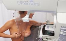
2. Mammography. Despite the fact that mammography is the most accurate method of breast examination, it still has some difficulties. In cases where the implant was installed above the pectoral muscle, during the examination it may block some areas of the breast. If the implant is installed under the muscle, the blocked area is insignificant. The disadvantage of this method is that it will not help determine whether a breast implant is ruptured or leaking. When performing mammography, each gland is compressed first horizontally, then vertically, so the patient should warn the doctor in advance about the presence of breast implants in the mammary glands.

3. MRI - magnetic resonance imaging. The peculiarity of the method is the use of a powerful magnetic field. MRI allows you to determine rupture or leakage of the implant, identify tumor foci and metastases.

4. CT - computed tomography. This type of examination is considered the most accurate for diagnosing breast cancer. Computed tomography is classified as an X-ray method for examining the breast.
5. Fluorography. Breast implants are visible in the fluorography image. The patient should notify the doctor in advance about their presence, but they will not interfere with the examination of the lungs, because implants easily transmit x-rays.
Thus, the presence of breast implants will not interfere with any of the listed types of examinations and obtaining accurate data on the condition of the patient’s mammary glands and lungs.
06.03.2017
The difference between natural breasts before surgery and after surgery to install implants is obvious
The implant can be placed subcutaneously, under the pectoral muscle fascia and behind the pectoralis major muscle. 
The artificial breast looks most natural in the latter case, but for the patient this is the most painful type of breast surgery. The implant stretches the muscle and the sensations are approximately the same as with a strong stretch of any other muscle.
This is what a breast implant looks like.

Selected by the patient according to the desired size.

One of the unobvious disadvantages of breast implants is back pain. The body does not immediately get used to excess weight in front of the chest.
A sign of artificial breasts will be the absence of prolapse of the mammary glands under clothing, especially with a large size.
On radiographs in direct projection, the implants look like this

The main danger from implants is being accidentally pierced or crushed. In this case, surgery to change the implant will be required.

When implanting, it is very important not to overdo it with the size so that the breasts look beautiful and not strange.

Patients say that after the operation it is easier to get married.

On a lateral radiograph

Breast implant on MRI

Breast implants on a thermal imager

Implant installation method

Implant in soft x-ray

Natural breast size 3

Natural female breast of the third size in an x-ray image - a positive image

And lastly - x-ray humor

FAQ:
1. Is it possible to do fluorography with silicone breasts? - Can! But the radiologist and x-ray technician find out that the breast is not natural. But they won’t tell anyone, because they are not interested.
Link to articles.
If a patient has had dental implants installed at some time, throughout his life he will have to follow certain recommendations for their use and care of the structure. There are certain medical procedures in which the presence of implants is necessarily taken into account. One of the most common questions is whether it is possible to do MRI with dental implants. It is immediately worth noting that before performing an MRI procedure, the doctor must be warned about the presence of prostheses in the oral cavity.
Major Limitations for MRI
So, is it possible to do an MRI with titanium implants? This is a common and quite important question that has become relevant with the development of modern medicine. MRI is a completely new innovative method of examination, during the use of which any pathological process can be detected, and at the very early stage of its development. Using this form of examination, you can examine any part of the body, including bone tissue and dental cartilage. If there are any problems with this part of the body, an MRI may be performed. With its help, you can determine the condition of the implants, whether there is an inflammatory process near them or not.
An MRI with a variety of dental structures may be ordered by a dentist to detect the following diseases and signs of discomfort. A decision is made to perform a magnetic examination in case of such unpleasant sensations as:

Thanks to a competently performed MRI procedure, it is possible to understand whether the patient needs surgical treatment, and whether or not simple drug therapy can be used.
Types of material for dental structures and MRI
The answer to the question whether or not it is possible to carry out resonance tomography with dental implants directly depends on which of the materials was used in the prosthetics process. Some time ago, structures of this type were made of metal, as a rule, an alloy of gold, copper and steel was used. This composition may not have a significant effect on MRI, but the picture that is obtained during the study is not very clear. Currently, implants made of metal ceramics are used in dentistry. With them you can undergo MRI without any problems, the quality of the image will not be spoiled at all.

If there is pain, the procedure may still need to be done.
Not only the material of the crown matters, but also the bases from which all the parts are made, that is, the pins and screws. Each such element is made of paramagnetic, ferromagnetic and diamagnetic material. Such bases are able to influence the magnetic field that is created by MRI machines. These are special materials that, under the influence of the magnetic field present in tomography equipment, can heat up and move. For this reason, before the examination it is worth checking the shape of the dental structures that stand, their strength and the reliability of their fastening.
Insufficient MRI result - reasons
Implants do not pose any danger during MRI. The only unpleasant point is a slight distortion of the results. For this reason, each patient who plans to have an MRI should notify the doctor. The procedure can be carried out without any problems, but to obtain the most accurate settings, the specialist will make preliminary adjustments to the equipment. It is strictly prohibited to conduct an examination in the following cases:

Another important rule that applies to people with dental implants is a preliminary x-ray examination. On them, the specialist will clearly see the clear location of the dental structures.
MRI and metal crowns
MRI of the jaw and teeth is prescribed in the rarest cases. The main reason is the fact that these areas of the body are quite difficult to access for research. The procedure is indispensable for various discomforts during chewing, spasms in the jaw area, as well as some restrictions in jaw mobility.
If you are wondering what to do if a crown or pin made of ferromagnetic alloys is installed. If such materials are present in the oral cavity, MRI is performed only with the permission of the attending physician. The specialist is already deciding what to do, whether the examination can be carried out or not. It is worth noting that in some modern clinics in large cities, MRI rooms are equipped with equipment that does not displace implants or heat them.

Metal crowns cause blurry images.
In a situation where the clinic does not have the appropriate equipment at its disposal, the procedure may be refused. There are fewer and fewer such medical centers; the majority of clinics are equipped with the most modern equipment. Professionals can set the settings in a special way and get a clear picture of the information equipment. In some particularly critical situations, when a person's life is at stake, the patient will be advised to remove all dental crowns.
Summing up
Modern magnetic resonance imaging is a special innovative method for diagnosing the human body. Despite the presence of a large number of positive factors and indications for the use of this examination, there are some contraindications. Among them, we can highlight the presence of implants. In such a situation, there is no need to worry about the structure, since it is not damaged under the influence of the magnetic field.
To avoid such a negative impact, it is worth notifying the doctor about this factor in advance. Having received such information, the professional will make a decision on how to proceed, how best to diagnose in order to prescribe high-quality effective treatment.
If all rules are followed and the doctor’s recommendations are followed, an MRI will be performed without any consequences for the teeth and the body as a whole. Based on the data obtained, the doctor develops a scheme for subsequent actions.
A safe method for studying pathologies of internal systems and organs is magnetic resonance imaging. This type of diagnostics is successfully used in all areas of medicine. Is it possible to do an MRI with dental implants? In certain cases, this may be a contraindication to the procedure.
The essence of the method
MRI is a modern and universal way to diagnose disorders of the human body. The essence of tomography is the use of such a physical phenomenon as nuclear magnetic resonance. During the study, hydrogen atoms located in the tissues of the human body emit weak radio signals. They are “caught” and organized by a special device - a tomograph, consisting of a magnet and maintaining a constant magnetic field. A computer helps process and convert the received information into a three-dimensional image.
Completely eliminates x-ray radiation. Therefore, most patients with certain indications can undergo MRI. The procedure is prescribed only on an individual basis. The magnetic resonance method is completely safe for the human body.
Indications for use
The range of diseases for which a patient can be prescribed an MRI is quite wide. Due to the absence of X-ray radiation, the method is used to study pregnant women and children from a very early age.
Is it possible to do an MRI with dental implants, braces or crowns? This method is an alternative that has more contraindications.

Most often, the technique is used when it is necessary to diagnose soft tissues and blood vessels. It is recommended to carry out diagnostics in the following cases:
- circulatory disorders;
- thrombosis;
- hemorrhages;
- vascular aneurysms;
- heart diseases;
- pathologies of the musculoskeletal system;
- inflammatory processes of various origins;
- diseases of internal organs.
Is it possible to do an MRI with dental implants?
A huge number of people now have dental structures and products for various purposes, but this is not a reason not to undergo medical diagnostics. In dental practice, magnetic resonance imaging makes it possible to assess the condition of the dentofacial elements that are visible and hidden for easy viewing. If a patient has pins and other metal structures in the oral cavity, can an MRI be done? Many people undergo this procedure with dental implants.

Dentists assure that the presence of metal elements is not a contraindication to magnetic resonance therapy. To get the correct result, you must first warn the specialist about the presence of metal elements and the type of material from which they are made.
When is an MRI prescribed in dentistry?
Patients suffering from the following pathological conditions require diagnostics:
- pain when chewing food;
- crunching when moving the jaws;
- pain syndrome in the lower jaw;
- spasm when closing and opening the mouth.
MRI of the brain
In neurology, magnetic resonance therapy is used to study dysfunction of the vessels of the brain, spinal cord, and spinal column. MRI of the brain with dental implants is indicated for increased intracranial pressure, suspected stroke, or skull injuries. Diagnostics will help determine the cause of frequent headaches, developmental pathologies, and the presence of infectious diseases.

A more detailed picture of the disease can be obtained by using contrast in the diagnostic process. To do this, the patient is injected with a special substance that enters the blood vessels of the brain through the bloodstream. When exposed to a magnetic field, problem areas in the image are colored.
Implant materials
The material from which the dental structure is made can be paramagnetic, ferromagnetic or diamagnetic. They behave differently when exposed to a constant magnetic field. Is MRI done with ferromagnetic alloys? It is quite possible to carry out the procedure, however, the final result may be greatly distorted.

Currently, experts give preference to paramagnetic alloys - non-magnetic alloys. One of the popular materials is titanium. It has a number of advantages compared to other alloys:
- no toxic effects;
- high rates of survival of titanium implants with tissue;
- high strength and ductility;
- absence of vanadium in the alloy;
- the presence of an oxide film on the surface that protects from the effects of the external environment;
- the material is not an allergen.
Features of the procedure
MRI with dental implants made of gold, platinum, palladium alloys can show a distorted picture of the disease. Conducting research in this case is not recommended. It should be borne in mind that the procedure will not cause any harm to a patient who has ferromagnetic implants. Although some people tend to think that movement or heating of the dental product may occur. Implants are small in size and well fixed, and therefore there is absolutely no need to be afraid of anything.
However, the resulting image will be unreliable and will not allow the doctor to make a correct diagnosis. Before the procedure, you should definitely check with your dentist about the type of material of the dental structures. In the future, this information must be communicated to the specialist who will conduct magnetic resonance imaging. This will allow you to correctly configure the equipment and get a true picture of the patient’s condition.
Magnetic resonance imaging is presented as an innovative examination method, thanks to which it is possible to diagnose any pathological processes, even if they are at an early stage of development. , since it allows you to study in detail the cartilage, bone structure of the teeth and the soft tissues that are adjacent to them. Against the background of this trend, a completely logical question arises: is it possible to do MRI with dental implants?
Dental implants are special posts that are implanted by a dentist into the lower or upper part of the jaw for later use as a support for dentures. The procedure is very common in modern dentistry, as it allows you to correct any dental defect. Returning to the question of the possibility of performing an MRI in the presence of dental implants, it is necessary to know what material was used to make the post, since the magnetic field of the tomograph affects different materials differently, which can be classified as paramagnetic, diamagnetic or ferromagnetic.
- Implants made of nickel, cobalt, iron and other similar materials are ferromagnetic and under the influence of a magnetic field they are actively magnetized, and most importantly, they interfere with obtaining a reliable result, leaving artifacts (interference) in the images.
- Products made from diamagnetic materials do not change their temperature under the influence of a magnetic field, since they repel the magnetic field. Examples of such materials include silver or gold.
- The most popular and widespread are implants made of paramagnetic materials, which are inert to the magnetic field. It can be zirconium, platinum or titanium.
Titanium implants take root well in the patient’s body. In modern dentistry, this material is most often used. The MRI procedure for patients with such implants in the gums takes place without any negative consequences. Before undergoing a diagnostic procedure, it is worth checking what material was used in the manufacture of your orthodontic appliances.
Patients' concerns about the possibility of performing an MRI in the presence of structures in the oral cavity are associated primarily with medical series, where during the diagnostic procedure the skin in the area is torn off, pins are pulled out of bones and teeth. In fact, magnetic resonance imaging does not lead to such consequences, especially if paramagnetic materials were used as materials for implants. They do not react in any way to the field created by the tomograph, so they do not cause negative consequences.

Myths and reality
Many people associate having an MRI with dental implants with dire consequences, but it’s time to debunk these myths.
- The first common myth is that implants become dislodged during the diagnostic procedure and damage soft tissue. In fact, the force of a magnetic field can only displace those elements that are not fixed in the body. These could be surgical clips on the vessels of the brain or on the heart, shrapnel, bullets. As for the pins, they are securely fixed in the bone tissue and will not be able to move and damage the oral cavity during an MRI.
- The second myth is related to the tomograph’s ability to heat metals. It was already mentioned earlier that the magnetic field actively affects only ferromagnets, that is, medical products made of steel. Depending on the dimensions of the product, they may indeed heat up during the diagnostic procedure, but you should only be wary if the dimensions of the product are large enough. Don't worry, there are no implants of this size in the oral cavity. As for other materials, they are absolutely safe and do not heat up during an MRI.
If the patient still has concerns about pain, there is no need to worry, since there is always the opportunity to use a special button that the specialist gives before starting the diagnosis.
How does the patient feel during the procedure?
Many patients with implants are worried about how they will feel during an MRI and whether there will be pain. However, there is no need to worry about this. The effect of a magnetic field on the body is determined only by the chemical composition of the alloy from which the implant is made, so this nuance must be clarified before the procedure. Otherwise, the operation of the tomograph is standard and does not cause pain or discomfort, with the exception of noise, which can be avoided using headphones.
Possible image distortion
Distortion of diagnostic results is the only unpleasant consequence of an MRI in the presence of dental implants, which is why the specialist must be warned about the presence of such implants. The presence of pins in the teeth is not a categorical contraindication; however, the results may be distorted, but this can be avoided.
On different tomograph models used by a particular clinic, a specialist can change the settings depending on the type of dental implant, which will make the images as clear as possible. To do this, the patient must take an X-ray of his jaws and bring a panoramic image, which will be performed by MRI. The photo should clearly show the dentures and their location. MRI may also be used to examine the oral cavity, but such situations are extremely rare, since traditionally this is done using conventional x-rays or computed tomography. When using a tomograph to scan other areas of the body, the results are not distorted, and the procedure proceeds in a standard manner.
As you can see, MRI with dental implants follows a standard procedure and does not cause negative consequences or pain. The only thing you need to do is to warn the specialist in advance about the presence of foreign bodies in the body and clarify what materials they are made of.


 Ponomarev I and the psychology of creative thinking republished")

Zaraisky D.A. Managing other people's behavior - file n1.doc. Food industry enterprises
Stanislav Müller holographic memory: books about the secrets of super memory What is family education
Robert Greene the art of seduction and seduction
Personnel accounting and payroll calculation in "1C:ERP" Document "Reflection of salaries in financial accounting"
Which account is used to record banking transactions?