Type Coelenterates(Coelenterata), as well as sponges, the oldest of the multicellular organisms, are known from the Vendian (Vendian - the last epoch of the Proterozoic), in the Ordovician of the Paleozoic they were already represented by numerous groups. Coelenterates are predominantly marine, solitary or colonial organisms, characterized by two life forms: attached polyp and free-floating jellyfish. In many coelenterates, both forms alternate during the life cycle ( metagenesis), some coelenterates (hydra, coral polyps) do not have jellyfish, others (certain species of scyphoid jellyfish) have lost the generation of polyps.
The body of an individual coelenterate consists of two layers of tissue - ectoderm And endoderm, between which there is a layer of gelatinous mesoglea. The ectoderm consists mainly of epithelial-muscular cells, combining integumentary and motor functions, from those characteristic of coelenterates stinging cells, forming stinging capsules (nematocysts), and undifferentiated cells, giving rise to cells of all types. In the endoderm, in addition to epithelial-muscular and stinging cells, there are glandular digestive cells. Intestinal cavity, or gastric cavity, simple or divided into chambers (in polyps) or canals (in jellyfish). Mouth, surrounded tentacles, serves to capture food, as well as to remove undigested residues. Digestion is cavity and intracellular. Nervous system diffuse type. Jellyfish, in addition, have two
Drawing. Stinging cells characteristic only of Coelenterates.
nervous rings and sense organs - either photosensitive eyes, or statocysts, and in scyphojellyfish - Rhopalia.
Reproduction is sexual and asexual. Incomplete asexual reproduction in a number of species leads to the formation of large colonies. Many coelenterates are dioecious, some are hermaphrodites. In hydroids, reproductive products develop in the ectoderm, and in scyphoid and coral polyps, in the endoderm, after which they are released into the external environment, where fertilization occurs. A free-swimming larva develops from a fertilized egg - planula. A polyp (less commonly a jellyfish) is formed as a result of metamorphosis of the planula. Jellyfish usually bud on the body of polyps. In some species, development occurs in the mother's body, and young individuals are excreted through the mouth.
There are about 9 thousand species of modern coelenterates and about 20 thousand extinct species. Coelenterates are found in all seas, from the surface to extreme depths and on the bottom. There are freshwater species (hydra). All coelenterate predators feed on plankton and larger aquatic organisms, some are food competitors of fish, and some serve as food for other organisms.
Type Coelenterates are divided into 3 classes.
Class 1. Hydroid polyps, or Hydrozoans (3 thousand species). The most famous among hydroid polyps is Hydra. This is a small (up to 1-3 cm) polyp found in our fresh water bodies. It leads a sessile lifestyle, attaching itself to the substrate with its base, or sole. At the free end of the body there is a mouth opening, surrounded by a corolla of 6-12 tentacles, on which the bulk of the stinging cells are located. Hydra feeds mainly on small crustaceans - daphnia and cyclops. Reproduction occurs by both sexual and asexual (budding) methods. In the first case, a new hydra develops from a fertilized egg after a period of rest (in winter).
However, most hydroid polyps, unlike hydra, lead not a solitary, but a colonial lifestyle. At the same time, in such colonies special mobile individuals arise and bud off - the very jellyfish that are responsible for the dispersal of polyps. Jellyfish actively move and release mature germ cells into the environment. The larva that has developed from a fertilized egg also moves in the water column for some time, and then sinks to the bottom and forms a new colony.
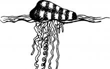
The class Hydroid polyps from the subclass Siphonophora includes very interesting colonial animals from the genus Physalia. These are marine organisms that live mainly in the southern seas. Although outwardly Physalia looks like a solitary animal, but is actually a colony of organisms. In this colony, individual individuals are attached to a single trunk, in which a common gastric cavity is formed, communicating with the gastric cavity of each individual. The upper end of the trunk is swollen, this swelling is called air bubble, or a sail, or a float. The air bubble is one highly modified medusoid individual. Along the edges of the hole leading into the cavity of the bladder, a closing muscle is formed: the bladder can release gas from the hole (it is secreted by the glandular cells of the bladder, its composition is close to air), and thanks to this, Physalia able to float to the surface or dive into the depths. Below the bladder there are other individuals specialized in feeding or reproduction, and there are also individuals with particularly abundant stinging tentacles. One of the most common physalia of the Pacific Ocean ( Physalia utriculus) one of the tentacles, the so-called lasso, longer than all the others, and can reach 13 or more meters in length. Along it are located thousands of stinging batteries, each of which consists of hundreds of microscopic stinging cells. When a fish encounters a tentacle, threads of stinging cells pierce the tissues of the victim, and the poison from the capsules is pumped through these channels. Thus, the lasso captures and paralyzes fairly large prey, and then pulls it towards the mouth.
If Physalia will sting a person who accidentally touches it, the consequences can be very serious. Burns Physalia are very painful, blisters appear on the victim’s skin, lymph glands become enlarged, sweating increases, nausea appears, and it becomes difficult for the person to breathe.
A close relative of Physalia is interesting - Portuguese warship(Physalia physalis). It is found in the tropical Atlantic, Caribbean and Mediterranean waters. Similar species of physalia live off the Hawaiian Islands and off the coast of southern Japan. Portuguese man of war got its name from its bright, multi-colored floating air bubble, reminiscent of the sail of a medieval Portuguese ship. Its ridged air bladder, approximately 35 cm long, is very colorful. The membrane of the bladder is colored iridescent blue, turning into mauve and then, at the top of the ridge, into pink (you have to imagine it). Boat colonies look like unusually elegant balls, often drifting in whole clusters on the surface of the ocean. From time to time, the boat dips its bladder in water so that the membrane does not dry out. Deadly poisonous tentacles stretch 10-15 m down from the bubble, capable of paralyzing even very large fish and pulling it up to the mouth opening. One researcher talked about his meeting with this cute boat: "...without thinking, I grabbed it, and roared in pain, frantically began to wash my fingers with sea water, but the sticky mucus did not lag behind. An attempt to wash off the mucus with soap was also unsuccessful. My hands burned and ached, my fingers bent with difficulty. Spraying with an anesthetic medicine from a special The atomizer relieved the pain for a few minutes, but it immediately returned with renewed vigor. The fingers could no longer bend, the pain began to spread to the shoulders and further to the heart area, the general state of health was disgusting. I took two tablets of analgin, validol, pyramidon, and, as they say, I fell into bed. I was shaking with chills... It subsided gradually. At first my right hand felt better, then my left one. The pain subsided only after five hours.

Drawing. Colonial hydroid polyps Physalia (with fish)
and Portuguese Man of War (right).
Although physalia are inhabitants of the open ocean, many of them, under appropriate currents and weather conditions, are carried to the shores of Northwestern Europe. Even washed ashore, they retain the ability to sting anyone who touches them. Sometimes Portuguese ships fall into the Gulf Stream and are carried by this current into the English Channel. When they accumulate off the coasts of England and France or, for example, near the beaches of Florida, television, radio and print warn the population of the danger.
Despite the toxicity of physalia, some sea turtles eat them in huge quantities. People, of course, do not eat physalia, but they also find uses for them. Their venom is unusually resistant to drying and freezing, and the tentacles, which lay in the refrigerator for six (!) years, perfectly retained their deadly properties. Farmers in Guadeloupe (Caribbean) and Colombia use dried physalia tentacles as rat poison.
Class 2. Scyphoid jellyfish, or Scyphozoa (200 species of different jellyfish). Scyphoid jellyfish are solitary, actively swimming inhabitants of the temperate and tropical waters of the World Ocean. The body of most jellyfish is transparent, which is due to the high (often up to 97.5%) water content in the tissues. In scyphoid jellyfish, the body has the shape of a rounded umbrella with long tentacles suspended from below. In all species, a gastrovascular system of varying complexity is formed: the oral opening, which is located on the lower side in the center of the umbrella, leads to a large stomach, from which the gastric canals diverge radially. A number of tentacles in jellyfish are modified, turning into so-called marginal bodies. Each of these bodies carries one statocyst (a formation involved in maintaining balance) and several ocelli, including some of a very complex structure.
Jellyfish go through two levels of development: sexual - this is the jellyfish itself, and asexual - this is the polyp. The common jellyfish reproduces sexually. Male reproductive products are released through the mouth into the water, after which they enter the female’s body, where fertilization occurs. The egg develops into a mobile larva - a planula, which is released into the water, goes to the bottom and attaches to underwater objects. Thus, it turns into a single polyp - scyphistoma. It grows, feeds, and then begins to reproduce by division (strobilation of scyphistoma). A mature polyp disintegrates into several discs, which turn into small jellyfish - ether. The ethers grow and turn into mature jellyfish.
In general, jellyfish are round like a ball, flat like a plate, elongated like a transparent airship. They can be quite small, such as Chironex, or sea wasp(no more than 3 cm in diameter), and huge, like a giant of the Arctic waters, fiery red Cyanea, or Lion's mane, whose domed body grows up to two and a half meters in diameter, and bundles of writhing thread-like tentacles reaching 30 m in length can cover a five-story building! (The maximum recorded length of the tentacles of the giant Arctic jellyfish Cyanea was 36.5 m, and the diameter of the dome was 2.3 m. It washed ashore in North America in 1870. This specimen was greater than the maximum length of the blue whale, which is considered the most largest animals on the planet.)

Drawing. Scheme of development of the Scyphoid jellyfish.
1 - egg, 2 - planula, 3 - scyphistoma, 4 - budding scyphistoma, 5 - strobilation, 6 - ether, 7 - adult jellyfish.
A much more modest jellyfish in size Pelagia, or Nochesvetka, amazes experienced sailors with a bright light in the middle of the night in the waters of the Mediterranean Sea, demonstrating the phenomenon of bioluminescence.
However, the beauty of most types of jellyfish can be very deceptive. After all, to a greater or lesser extent, all jellyfish are poisonous. The only difference is that some species are practically not dangerous to humans, others sting like nettles, and a painful burning sensation can be felt for several days, and others cause paralysis that can lead to death.
There are also jellyfish that are completely harmless to humans. This is the well-known glassy-white “eared” jellyfish - Aurelia. It lives in all tropical and moderately warm seas, including here in the Black Sea. Aurelia reaches 40 cm in diameter. Umbrella Aurelia translucent, most often colorless, sometimes there are umbrellas with a slight shade of blue, pink, purple. Aurelia- these are the animals of the summer season. Autumn storms bring death to them, so on the eve of cold weather, small, slightly more than a centimeter, lumps of living tissue settle to the bottom of the sea Aurelia, which carry hereditary information about this organism. These lumps are not afraid of either storms or cold snaps, and with the arrival of spring, tiny discs separate from them, which grow into adults in one summer. By the way, if you rub Aurelia’s body into human skin, it becomes immune to “stinging” jellyfish, such as, for example, the same Black Sea Rosistoma, or, in other words - Cornerot.

Drawing. Jellyfish Cyanea (A) and Jellyfish Aurelia aurita (B).
Cornerota can be recognized by the large size of the umbrella, up to 50 cm in diameter, and large fleshy root-like outgrowths. But these are not tentacles. U Cornerota there are no tentacles, their oral lobes branch, forming numerous folds fused together. The ends of the oral lobes do not form folds, but end in root-like outgrowths.
Cornerot- a predator that prefers small fish, worms, and small crustaceans. With its poison, it paralyzes prey and successfully eats it. However, mackerel fry travel in flocks together with the cornet jellyfish - they are not afraid of the stinging glands. But protection is an absolute plus of such a symbiosis.
In the Black Sea Cornerot widely spread. A particularly large number of individuals of this species appear on the coast in the second half of summer. This is far from the most pleasant part of the holiday, but it’s not dangerous either: the poison of one Cornerota is not fatal to humans, and the pain after a burn is not much stronger than nettle. Cornerot sensitive to changing weather conditions. For example, before a storm, jellyfish move away from the shore and go to the bottom.

Drawing. Black Sea jellyfish Cornerot.
Jellyfish Gonionema- a real baby among the whole multitude of sea jellyfish. Its size is no larger than a coin (3-4 cm in diameter), and its body has the shape of a flattened bell, along the edges of which there are many tentacles with suction cups, sometimes up to 70-80 pieces. Gonionema has a dome with four brown folds in the form of a cross on the concave side. This is why they called the jellyfish cross. Despite its small size, this baby is Jellyfish-cross- in many ways even more dangerous than its larger brothers. It lives in the waters of the Pacific Ocean: in the Sea of Japan - near Vladivostok, in the Tatar Strait, near the southern outskirts of Sakhalin, off the coast of Japan and the South Kuril Islands.
It lives in shallow water in algae thickets, so encounters with people are not uncommon. The danger of these jellyfish is not that their poison is particularly toxic, but that they live in shallow water, and their invasions are sometimes spontaneous. For example, in July 1966, a huge flock of Krestovichkov- thousands of vacationers were affected. There, in the summer of 1970, they received burns from Krestovichkov 1360 people, of which 116 had to be urgently hospitalized.
The poisonous apparatus of these jellyfish is located in the outer layer of the tentacles. These are chitinous capsules filled with poison. Each cell has a sensitive hair or the thinnest tube, the so-called stinging filament. Any touch causes a reflex release of a portion of poison. In case of accidental contact with Cross in water it is not so easy to get rid of it: as if afraid of losing its prey, it is firmly attached to the body. It has to be torn off with force.
Of course, it is impossible to die from touching one jellyfish, but the sensations are far from pleasant: at first you feel a tingling sensation, similar to those you get from touching nettles, but unlike a nettle burn, meeting with Cross entails serious consequences. Swelling, rash, burning sensation, itching appear at the site of the lesion; sharp pain in the lower back and joints, shortness of breath, dry cough, nausea, numbness of the arms and legs. Sometimes there are convulsions. I Krestovichka often affects even the psyche. Usually, poor health lasts 4-6 days, but pain and discomfort may recur for about a month. Repeated meetings with Cross extremely dangerous. The whole point is that the human body does not develop immunity to its poison, but becomes even more sensitive to it.
It's most dangerous when Crosses attack in packs. The poisoning of the body is so great that instant death can occur. To avoid burns Krestovichkov you need to stay away from the algae thickets where these jellyfish live. When working near corals, do not touch them with your bare hands.

Drawing. Medusa-cross.
The most dangerous of all existing jellyfish - Sea wasps. They live in the warm waters of the Indian and Pacific oceans. It's hard to believe that this little blob of living mucus is actually a real killer. And meeting him is almost more dangerous than meeting a shark. I sea wasp so strong that if it enters the bloodstream, it can stop a person’s heart in a few minutes. In search of food, such as bottom-dwelling shrimp, these deadly creatures sometimes come very close to the shore. Large concentrations of small larvae are often observed in the coastal waters of Australia and the Philippine Islands Sea wasps, the local name is “stinging sea grass”, or “stinging pine needles”. Once caught in such a congestion, a person can suffer severe burns if his body is not protected by clothing.

Drawing. Jellyfish Sea wasp.
In the waters of the Sea of Japan, the polypoid stage of jellyfish Navzitoi reefs and rocks are covered with continuous poisonous thickets. The Japanese call this polyp "iramo", which means stinging algae. It’s not for nothing that local fishermen and divers are afraid of such places. But some other slightly poisonous species of jellyfish are considered an exquisite delicacy in Japan and, after special processing, end up on the table... fried! This very exotic delicacy requires special cunning in preparation, especially considering that jellyfish consists of 90 percent sea water.
As you can see, an encounter with jellyfish can have very unpleasant consequences. Treatment required. During treatment, it is necessary to reduce pain, reduce spastic (convulsive) phenomena and eliminate local lesions (burns). It is recommended to administer analgesics to reduce pain. For local treatment, lotions with diluted ammonia, ethyl alcohol, and oil compresses are used. If cardiac or respiratory disorders develop, symptomatic treatment should be applied. Along with medications, it is advisable to use heat (heating pads, hot tea, rubbing hands and feet, etc.). For skin rashes, antihistamines must be administered.
Prevention consists of avoiding contact with poisonous jellyfish and siphonophores. During emergency underwater work in areas where these animals live, it is necessary to wear fairly thick clothing (wetsuits) and gloves. If there are large concentrations of small jellyfish, you should protect your eyes. In the event of a burn, the victim must get to shore or on board the ship as soon as possible. There are cases where, as a result of burns, people lost consciousness from pain and drowned before help came to them.
Fishermen engaged in commercial fishing may come into contact with jellyfish when retrieving nets, disassembling the catch, and processing fish in production facilities.
The gelatinous body of a jellyfish, consisting almost entirely of water, is easily destroyed, and therefore whole specimens are not always preserved in the catch, by which one can determine whether a given jellyfish is dangerous or harmless. Therefore, any jellyfish that comes on board a vessel should be handled with care. Pieces of burning tentacles can stick to nets and ropes when hauling gear on board the vessel and, together with splashes of water, get on the face and, which is especially dangerous, in the eyes. Therefore, when working in habitats of poisonous jellyfish, it is necessary to use gloves (mittens) and safety glasses. Remains of jellyfish should be removed (washed off) from the deck and gear, since, once dry, they can get into the eyes in the form of fine dust and cause dangerous inflammation.
Class 3. Coral polyps(6 thousand species). Coral polyps (Anthozoa) are colonial (less often solitary) marine organisms. The body ranges in length from several millimeters to one meter and has six-ray or eight-ray symmetry. Due to the fact that fertilization in corals is internal, the planula larva develops in the gastric cavity of the polyp, which forms eggs. There is no jellyfish stage. The oral opening is connected to the gastric cavity by the pharynx. Polyps of one colony have a common gastric cavity, and food obtained by one of the polyps becomes the property of the entire colony.
There are about 6,000 species of coral polyps; they live in all seas with fairly high salinity; There are about 150 species in the northern and Far Eastern seas of Russia.
Madreporaceae, or reef-building corals (from the group of six-rayed corals) surround themselves with a massive calcareous skeleton. When a polyp dies, its skeleton remains and, growing over thousands of years, forms coral reefs and entire islands. Madrepore corals- These are reef-building corals. The largest existing reef, the Great Barrier Reef, stretches along the eastern coast of Australia for 2,300 km; its width ranges from 2 to 150 km.
The skeleton of madrepore corals is quite complex. It is built by the cells of the outer layer (ectoderm) of the polyp. At first, the skeleton looks like a small cup-like cell in which the polyp itself sits. Then, as radial partitions grow and form, the living organism finds itself, as it were, impaled on its skeleton. Colonies Madrepore corals are formed as a result of budding. Some corals have not one, but two or three polyps in each cell. In this case, the cell stretches out, becomes like a boat, and the mouths are arranged in one row, surrounded by a common rim of tentacles. In other species, dozens of polyps are already sitting in the limestone house. Finally, in corals of the genus Meandrins all polyps merge to form a single organism. The colony takes on the appearance of a hemisphere covered with numerous winding grooves. Such corals are called brain corals; the grooves on them are fused mouth slits lined with rows of tentacles.
Colonies of coral polyps grow quite quickly - branched forms, under favorable conditions, grow up to 20-30 cm per year. Having reached low tide, the tops of coral reefs stop growing and die, and the entire colony continues to grow from the sides. New colonies can grow from broken branches.
In order for coral polyps to grow calmly and build reefs, they need certain conditions. In shallow, well-heated lagoons, they can withstand water heating up to 35 °C and a certain increase in salinity. However, cooling water below 20.5 °C and even short-term desalination have a detrimental effect on them. Therefore, in cold and temperate waters, as well as where large rivers flow into the sea, coral reefs do not develop.
Coral reefs are unique ecosystems in which a huge number of other animals find shelter: mollusks, worms, echinoderms, fish. During the pre-glacial period, coral reefs fringed many islands. As sea levels began to rise, the polyps built up their reefs at an average rate of a centimeter per year. Gradually, the island itself disappeared under water, and in its place a shallow lagoon surrounded by reefs formed. The wind carried plant seeds to the reef. Then animals appeared, and the island turned into a coral atoll.
Coral reefs have existed since ancient geological eras, and more than 5,000 species of fossil corals have been described. Remains of corals were found in the Urals in deposits of the Cretaceous period (about 100 million years ago) and in the Moscow region (deposits more than 300 million years old). Fossil corals are reliable indicators of the age of sedimentary rocks. Many of them are associated with deposits of certain geological rocks, in particular coal. The discovery of such corals indicates the presence of this mineral in this place. For example, coal was discovered in the Donetsk basin.
By studying the structure of fossil corals, you can calculate the number of days in a year in different eras. The fact is that the walls of the calcareous tubes forming the skeleton of the colony grew in layers: their growth occurred only during the day and strictly obeyed the lunar (that is, ebb and flow) cycles. In addition, the annual growth rings also differ - dark stripes correspond to the winter season, light stripes correspond to the summer season. The width of the stripes depends on daily changes in illumination and water temperature. Analyzing the nature of the growth of tubes on the thinnest saw cuts, scientists calculated that, for example, in the Devonian period (about 400 million years ago), a calendar year, equal to the period of the Earth’s revolution around the Sun, lasted about 400 days - a day then was less than 22 hours. After 150 million years, the year already had 390 days. There is a gradual slowdown in the speed of rotation of the Earth around its axis.
Building stone containing coral remains is successfully used as a decorative material for interior and exterior decoration. This stone was once a shell rock and consisted of sedimentary rock, corals and mollusk shells. After hundreds of millions of years, it turned into solid rock. On its polished surface, corals form an intricate pattern, sometimes containing layers of different minerals. The fossils embedded in the stone give it a wavy texture. Such a structure has, for example, marble from deposits near Nizhny Tagil in the Urals.
Red noble coral Mediterranean Sea (Corallium rubrum) belongs to the eight-rayed corals and is not capable of forming reefs. Its colonies grow on the coastal slopes of the Mediterranean Sea at a depth of more than 20 m (usually from 50 to 150 m). Even in ancient times, divers used a special hook to extract corals from great depths. About the same Noble red coral, which has long been used for making jewelry, is still mined today.
Sea anemones, or Sea anemones- These are non-skeletal solitary coral polyps. Sea anemones coexist with hermit crabs, settling on their shells. Cancer is protected by stinging cells Sea anemones, and in return suffers cancer Sea anemone from place to place - to more favorable places for hunting. Other types Actinium cohabitates with a clown fish. Bright fish, immune to tentacle poison Sea anemones, attracts enemies, and sea anemone grabs them and eats them. Something goes to the clown too. Separate Sea anemones live (in aquariums) up to 50–80 years.
DRAWINGS THAT SHOULD BE DONE IN THE ALBUM
(6 pictures in total)
Lesson topic: Sponge Type –Spongia
Type: Sponges
Class: Ordinary sponges
Order: Siliceous sponges Genus: Badyaga – Spongilla

Rice. 1. Badyaga. External building.
1-colony
2-substrate
Lesson topic: Sponge Type –Spongia
Type: Sponges
Class: Lime sponges
Genus: Sikon - Sycon

Rice. 2. The structure of a single Sicon sponge.
1-sole
3-osculum
4-outer layer of pinacocyte cells
5-inner layer of choanocyte cells
6-mesoglea
7-paragastric cavity
→ - direction of water flow
Lesson topic: Sponge Type –Spongia

Rice. 3. Morphological types of sponges.
Lesson topic: Sponge Type –Spongia

1-pinacocytes
2-collencytes
3-choanocytes
4-scleroblasts
5-spicule
6-amebocytes
7-ovum
8-mesoglea
Lesson topic: Type Coelenterates -Coelenterata
Type: Coelenterates
Class: Hydroid
Squad: Hydras
Type: Hydra - Hydra sp.

Rice. 5. Hydra stalked. External building.
2-tentacles
3-sole
5 mouth opening
6-substrate
Lesson topic: Type Coelenterates -Coelenterata

1-ectoderm
2-endoderm
3-support plate
4-gastric cavity
The hydroid jellyfish belongs to the class of hydroids and coelenterates. The habitat is water. They are close relatives of polyps, but are a little more complicated. This type of jellyfish differs from others in that it can live forever, since the hydroid can regenerate from an adult to a child’s organism.
Jellyfish do not have a mouth, but they do have an oral proboscis. She can always trigger the revival mechanism. Fernando Boero reported about the degeneration of the jellyfish; while studying hydroids, he conducted experiments on them. He placed some of them in the aquarium, but, unfortunately, the experiment was disrupted, as a result of which the water dried up and Fernando discovered that the jellyfish did not die, but only threw off their tentacles, transforming into larvae.
Nutritional resources and eating process
Plankton, Artemia
The main resource in the food of hydroid jellyfish is plankton. For them, the basis of nutrition is Artemia, such jellyfish are considered predators. The tools for obtaining food are the tentacles, which are located on the edge of the umbrella body. The digestive system of these jellyfish is called gastrovascular. Jellyfish catch prey by passively moving their tentacles in the water, into which plankton falls, after which it begins active swimming. In such jellyfish, the nervous system consists of cellular networks that form 2 rings, one of them is the outer one, which is responsible for sensitivity, and the inner one is responsible for movement.
One of the hydroid jellyfish have light-sensitive eyes, which are located in the center of the tentacle. Hydra, by its nature, is a predator for food; it chooses ciliates, planktonic crustaceans, and also fry. They wait for prey by clinging to an aquatic plant and at the same time open their tentacles wide. When at least one tentacle reaches the prey, then all the other tentacles completely envelop the victim. And it quickly swallows its prey whole; when the hydra is satiated, its tentacles contract.
Reproduction
 Reproduction of hydroid jellyfish is more often external than internal. Mature germ cells move outward, after which blastula is formed and some of the cells end up inside, forming endoderm. After some time, several cells degenerate to form a cavity. After this, the egg turns into larvae - a planula, and then into a hydropolyp, which buds into other polyps, as well as small jellyfish. After which the little ones grow up over time and begin to develop independently.
Hydra is one of the most convenient objects for conducting experiments, with the help of which scientists studying regeneration in animals. When the hydra is cut in half, after some time it itself restores the missing parts. Also, this type of surgery is easy to perform without anesthesia and there is no need to use special instruments. Hydra has the property of restoring not only from half, but even from the smallest pieces many polyps are revived.
Hydra habitats
Hydroid jellyfish are not always found, but in large concentrations transported by currents. The benthic class includes stages of polyps that lead a sedentary life, the exception to which is class of planktonic hydroid polyps. Hydroid species are also capable of grouping with the help of the wind into huge groups, but hydroid polyps, when clustered, seem to be one whole. If the jellyfish and the polyp are hungry, their movement will be aimed only at obtaining food, but when the body is saturated, their tentacles will begin to contract and be pulled towards the body.
Habitat zones
Jellyfish move depending on the presence or absence of hunger. In general, all species occupy a specific habitat, this can be either a lake or an ocean. They do not deliberately seize new territories for themselves. Alone prefer to live in warmth, while others, on the contrary, are in the cold. They can also be located both below at depth and on the surface of the water. Hydroid jellyfish can be found in the littoral zone, and they do not have a fear of the surf. Most of these jellyfish have a polyp, which is protected from impact by a skeletal cup (theca). The structure of the theca is thicker than that of other species that live deeper, where the perceptibility of the wave is much less.
At greater depths, a special type of hydroids lives, which is unlike littoral hydroids. At this depth there are colonies, having the form such as:
- tree,
- Christmas tree,
- feather,
- and there are also types of colonies that look like ruff.
Such species grow from 15 to 20 cm and cover the entire seabed with dense forest. Some species, for example, like the sea spider, live in these forests and eat hydropolyps.
Hydra can very rarely live in less saline waters, such as in the Gulf of Finland for such species, the salinity of the inhabited space should not exceed 0.5%. The hydroid jellyfish often lives close to the shore and in brighter places. This type of jellyfish does not have a tendency to be mobile; they most often attached to a plant branch or rock. One of the most favorite states of the hydroid jellyfish is to be upside down and have some tentacles hanging down.
Dangerous types of jellyfish for humans
 But not all can be safe for human life. One of the most beautiful species called "Portuguese man-of-war" may cause harm to humans. The bell, which is present in it and has a beautiful appearance, attracting attention, can cause harm.
Physalia, which is found in Australia, as well as on the coasts of the Indian and Pacific Oceans and even the Mediterranean, is one of the huge hydroid species. Physalia's bubble can reach a length of 15 to 20 cm. But Physalia's tentacles can be much scarier, since their length and depth can extend to thirty meters. Physalia can leave burns on the victim's body. An encounter with a Portuguese man-of-war is especially harmful for people with weakened immune systems and people prone to allergies.
But most hydroid jellyfish will not harm humans, unlike scyphoids. There is a so-called white algae from the genus polyps, which was previously used as decorative jewelry. Some of the hydroid species act as laboratory animals - these are polyps from the Hydra class, which are even used in schools around the world.
- a class of cnidarians whose life cycle includes a jellyfish with a characteristic feature - velum, and a polyp, which, unlike other cnidarians, never has internal partitions (septa) and a pronounced pharynx.
general characteristics
The life cycle may not have a polyp or jellyfish stage, but necessarily includes a planula larva. The lifestyle can be solitary (hydra) or colonial (obelia); in most species, colonies are formed in the polyp stages; There are colonies in which both polyps and jellyfish are integrated simultaneously (series Siphonophora).
Evolution
Fossil remains of hydroids have been known since the Precambrian; however, given the small number of solid skeletal structures, these remains are rather few in number and fragmentary. Recent studies of the structure of the medusar nodule (a special structure on a polyp that forms young jellyfish through specific budding) have given results indicating the presence of three germ layers in hydroids (i.e., hydroids are three-layered). The hydromedusa's pidparasol cavity and the layer of striatal muscle that lines it are formed from a morphological structure very similar to the schizocoel: in this case, a third layer (analogous to mesoderm) is formed between the ectoderm and endoderm, which turns into a cavity. Thus, the parasolic cavity is actually a coelom, which subsequently, after the formation of the velar foramen, becomes open to the outside.
There is also now available molecular biological data that shows that the genes that encode the formation of mesoderm structures in bilaterally symmetrical animals (Bilateria) are also present in hydroids. Thus, the polyp stage of hydroids has two germ layers (that is, two-layered), and the medusa stage has three germ layers (that is, three-layered). If these data are confirmed by evidence from other sources, this will mean that the transition from two-layered to three-layered organisms occurs every time a jellyfish buds from a polyp, and thus one of the biggest questions of animal evolution will be solved - how did it happen? transition from Diploblasta (animals with two germ layers) to Tryploblasta (animals with three germ leaves).
Taxonomy
Hydroids are known taxonomists from the very beginning of the existence of zoology as such; a large number of species were described by Carl Linnaeus back in the 18th century.
The taxonomy of hydroids is quite complex, which is caused by the lack of paleontological information on the basis of which it is possible to identify related relationships within the taxon. There are now several general classification options; in this article, the classification is based on the principles generally outlined on The Hydrozoa Directory website, and is largely based on the results of molecular biological research in recent years. According to the results of the mentioned studies, the hydroid class is clearly divided into two groups of series that have received the status of subclasses: Trachylinae and Leptolinae (the latter is also called Hydroidolina and Hydoidomedusae in other sources).
Hydroids is a cosmopolitan taxon, that is, one that is distributed throughout the world. They are found in both fresh and salt water.
Lifestyle
The jellyfish stage of the hydroid life cycle, as well as the polyp stage in the siphonophore, are mostly planktonic organisms. They occur seasonally, often in large aggregations that are carried by currents. Some jellyfish and siphonophores, however, are benthic. Stage polyps usually belong to the benthic, and lead a sedentary lifestyle, but there are exceptions: several planktonic hydroid polyps are known. In particular, the so-called swallowtail jellyfish is a planktonic free-swimming polyp. (Velella velella). The well-known Portuguese man-of-war is also a free-swimming colony formed from specialized hydroid polyps.
Most hydroids are predators and use the peculiarities of their lifestyle to catch prey. Planktonic stages transported by the current are often also capable of active movement in search of food. The location of the attached forms is determined by where the planula settles. Polyp colonies usually occur in areas where there is a constant flow of water, increasing the supply of potential food.
Behavior
Jellyfish lead a strictly individual lifestyle; they can be driven by the current into large aggregations, but so far no forms of social behavior have been recorded in them. Colonies of hydroid polyps, especially polymorphic ones, can be comparable to a single organism in terms of the level of specialization of individual polyps and the coordination of their actions. Polyps in a colony are usually descendants of a single planula, and are thus combined clones of identical genotype. However, in some species, colonies can mix their tissues or descendants of several planulae to form a single colony. In these cases, different individuals of polyps are in such a close relationship, forming (at the functional, but not at the genetic level) a single organism, which is probably one of the closest forms of social organization.
Most hydroids are dioecious. Fertilization is usually internal, without copulation. Males release sperm into the water by actively swimming past eggs attached to the mother's body (jellyfish or polyp) or thrown into the water by a female. Hydroids are the first organisms in which the presence of sperm attractors (substances that lure sperm during their free movement) was demonstrated, which provide species-specific attraction of sperm to eggs.
Members of the same colony of polyps (zooids) resort to coordinated behavior, requiring some communication between them. In such types as, for example, Thecocodium brieni, dactylozooids catch prey with their tentacles, while gastrozooids, after capturing prey, stretch to dactylozooids, remove the prey from their tentacles and swallow it. This division of labor, which includes advanced coordination, is quite common for polymorphic colonies.
Obviously, planktonic organisms cannot exhibit strong territorial behavior; but, as a number of studies have shown, free-swimming stages in the life cycle of hydroids actively avoid too dense aggregation of individuals of their species when feeding. Territorial behavior is pronounced among benthic organisms, where competition for suitable habitats is usually high. Thus, the high concentration of stinging dactylozooids on the periphery of the colony (in colonial species) is a protective adaptation aimed at limiting the growth of surrounding animals. In the same colonies, gastrozooids are capable of eating settling planulae of other species, and during development they can compete.
Both jellyfish and polyps, in a hungry state, continuously move in search of food; when the digestive cavity (coelenteron) is filled, the tentacles naturally contract and are pulled towards the body, which provides a certain degree of control over the rational expenditure of stinging cells (cnidocitives). The feeding behavior of many species of jellyfish leads to their periodic vertical migrations.
Nutrition
The main food resource of hydroids is plankton - in particular, small crustaceans. In laboratory conditions, the basis of nutrition for hydroids is, of course, artemia. Hydroid jellyfish are, for the most part, strict predators, and, in the case of feeding on fish eggs and larvae, can be considered the top of the food pyramid.
The diet of polyps is varied; Some species have symbiotic unicellular algae, and for a time they feed exclusively on the nutrients they supply during photosynthesis. Thus, these species can be considered functionally photosynthetic animals.
Forms of catching prey by jellyfish vary from passive hovering in the water column with motionless tentacles, which can be encountered by edible plankton, to active swimming in search of food objects. Polyps are capable of extending their tentacles and moving them to catch passing prey, but they can also resort to targeted hunting, which is provided by the sensory organs available (not in all species) that signal the approach of prey.
The main hunting weapon in hydroids is cnidocytes. Hydroids share a wide range of types of these stinging cells in all cnidarians.
From an ecological point of view, hydroid jellyfish, which feed on fish eggs, are the most dangerous predators for them; and the ability of polyps to feed on almost any larvae of fish and crustaceans includes them as an important link in the life cycle of a huge number of species. Thus, the ecological significance of the food specialization of hydroids is very great.
Reproduction
In hydroids, no signs of special mating behavior were found.
The eggs are stored in the gonads (gonophores) of females. According to the species, the eggs can be small and numerous, or large and few, up to one large egg per Gonophora.
The hydroid planula is, in fact, an embryo, not a larva, due to its extremely simple structure (in fact, it is a gastrula). Hydroid planula can be hollow (i.e. coeloblastula) or without an internal cavity (i.e. stereogastrula) of course, species that have a jellyfish in their life cycle are inherent in the hollow planula, which spends part of its life in the water column, swimming with the help of epithelial cilia. Species in which the medusoid stage is absent in the life cycle, of course, produce planulae without an internal cavity and immediately settle to the bottom next to the maternal organism (or colony). If there is a jellyfish in the life cycle, it is this generation that is “sexual,” that is, capable of sexual reproduction. The polyp generation is thus a specialized and perennial larva that produces a large number of sexual individuals during its existence. In many species, however, the medusoid stage can be partially or even completely reduced, and in this case the larva (Polyp stage), thanks to paedomorphosis, becomes a sexually mature individual. Almost half of the species of the subclass Leptolinae are characterized by a reduced or absent medusoid stage; thus, this group is a taxon with a common paedomorphosis of all animals.
Some jellyfish (for example, the genus Eleutheria) They have special brood pockets where they contain small young jellyfish. Also, some hydroids are characterized by gonothecae with brood chambers, which contain planulae for some time.
Many hydroid species are strictly seasonal, being active only for a certain period of time. Jellyfish can be observed for weeks or months, after which they completely disappear from the water column, and the species is represented by corresponding polyps in the benthos throughout the rest of the year. Polyps of the colony, in turn, can regress after waiting out a long period of hydrorhiza, reactivating when favorable living conditions return. Planula can encyst and wait out unfavorable conditions, similar to hydrorhiza, being covered with a protective chitinous shell.
Security status
There is not a single species of the hydroid class on the IUCN Red List. For most species, the exact boundaries of their range and abundance are unknown. A large number of species are considered endemic simply because they have not been specifically searched for outside the area of their initial discovery.
In the regional and national Red Data Books there are such representatives of hydroids as calcificans, coral-like families Milliporidae and Stylasteridae, which are also listed as CITES species. These families are traded, along with some other hydroids (known in the North Sea as "white algae"). The decline in their numbers is mainly due to habitat destruction.
Two species of this class are listed in the Red Book of Ukraine: olindias unexpected (Olindias inexpectata) and Merizia Azov (Moerisia maeotica).
Meaning for humans
Tremblay's famous treatise, which describes the transformations of hydroids of the genus Hydra, inspired Mary Shelley to write the novel Frankenstein; modern composer Frank Zappa wrote a song about hydromedusas, named by zoologists in his honor - Phialella zappai. But, of course, hydroids do not pay much attention to people.
"White algae" (colonies of polyps of the genera Hydrallmania And Sertullaria) were previously used as decorative ornaments until the populations of these hydroids began to decline catastrophically. Some hydroids are used as laboratory animals: the classic example is the polyps of the genus Hydra, which, along with scientific research, are used in school teaching in many countries around the world; but hydra is not the only example of such use: it is also widely used in scientific work Aequorea victoria(to obtain the marker protein aequorin), and species from the genera Hydractinia, Laomedea And Tubularia.
Some species of jellyfish can cause serious burns to humans; This danger also exists when coming into contact with polyp colonies of species such as fire corals (Millepora). When moving in large herds, even small jellyfish, such as members of the genus Cytia, can cause significant burns to swimmers.
But it is the feeding of some jellyfish that causes the greatest harm to humans (for example Aeroquorea Victoria) and free-floating colonies of polyps (such as Cytia gracilis) larvae and eggs of commercial fish.
Marine, less often freshwater animals that lead an attached lifestyle or swim in water. Attached forms are called polyps, floating - jellyfish.
Double layer animals, their body consists of two cellular layers: outer - ectoderm and internal - endoderm. Endoderm forms intestinal, or gastric cavity. The gastric cavity communicates with the environment through an opening that functions as oral And anal. Between the ectoderm and endoderm is mesoglea. In polyps, the mesoglea forms a supporting plate, while in jellyfish it forms a thick gelatinous layer.
Ectoderm cells perform protective and motor functions. The ectoderm contains special stinging cells that serve for defense and attack. Endoderm cells line the gastric cavity and perform mainly a digestive function. Digestion intracellular And cavity.
Breathing occurs through the entire surface of the body.
Nervous system absent-minded, or diffuse, type. Available tactile sensitivity, and in jellyfish, due to their swimming lifestyle, they are light-perceiving "eyes" And balance organs.
Coelenterates have radial, or radial, symmetry.
Asexual reproduction budding. Genital organs presented gonads. Fertilization is external. Some representatives are characterized by alternating asexual (polyp) and sexual (jellyfish) generations in the life cycle.
The type of coelenterates includes the following classes: Hydrozoans, Scyphoid jellyfish, Coral polyps.
Class Hydrozoa
Freshwater hydra
A BRIEF DESCRIPTION OF
|
Habitat |
Freshwater bilayer animals. Lead an attached lifestyle |
|
Appearance |
Saccular up to 1.5 cm. Radial symmetry. The mouth at the anterior end of the body is surrounded by tentacles, the sole is the posterior end of the body, for attachment |
|
Body cover |
Ectoderm - outer layer, endoderm - inner layer, mesoglea - middle layer |
|
Body cavity |
There is no body cavity. There is only an intestinal cavity |
|
Digestive system |
Blindly closed intestinal cavity. Mouth opening for food intake and for expelling undigested food debris. Digestion intracavitary and intracellular |
|
excretorysystem |
Ectoderm cells |
|
Nervous system |
Star-type nerve cells. Diffuse nervous system |
|
Sense organs |
Not developed |
|
Respiratory system |
None. Breathing through the entire surface of the body |
|
Reproduction |
Asexual - by budding. Hermaphrodites. Cross fertilization. |
GENERAL CHARACTERISTICS
This class includes small forms of coelenterates. Polyps And jellyfish belonging to this class are called hydroid.
Structure . The body of the hydra is oblong double layer bag, attached by the base, or sole, to the substrate (Fig. 1). Outer layer - ectoderm, inner layer - endoderm. There is space between the layers - mesoglea.
At the free end of the body there is oral cone, surrounded by a rim of 6-12 tentacles. Located on the oral cone mouth, employee and anus. The entire body surface is covered ectoderm, consisting mainly of cylindrical or cuboidal epithelial cells. Their base is extended upward and downward, along the longitudinal axis of the body, into a long process. The cytoplasm of the process differentiates as contractile fibers, in connection with this the process plays muscular role. The cylindrical parts of the cells form single layer epithelium. Thus, the cells perform a double function - cover And motor and are called epithelial-muscular. With the simultaneous contraction of all muscular processes, the body of the hydra is shortened. Between the epithelial-muscle cells there are small intermediate cells who participate in the formation stinging And germ cells, and also in the process regeneration- restoration of lost body parts or organs. Directly under the epithelium are located star-shaped nerve cells. Connected by their processes, nerve cells form the nervous system absent-minded, or diffuse, type. Of particular importance in the ectoderm are stinging cells, or capsules, serving for attack and defense.
Endoderm lines the whole gastric, or digestive cavity. The basis of endoderm cells is epithelial-muscular digestive cells. The muscular processes of these cells, unlike ectodermal ones, are located transversely with respect to the longitudinal axis of the body. When they contract, the hydra's body narrows and becomes thinner. Endodermal cells include glandular cells, secreting digestive enzymes into the gastric cavity, and cells with phagocytic activity. The latter are capable of capturing food particles using the movement of 1-3 flagella and the formation of pseudopodia. Thus, the hydra combines two types of digestion: intracellular And cavitary.
Rice. 1.The structure of freshwater hydra: a - longitudinal section; b - cross section; c - two-layer body; d - epithelial muscle cell; d - tentacle with discarded stinging threads; f, g - stinging cells; 1 - tentacles; 2 - testis; 3 - sperm; 4 - gastric cavity; 5 - budding young hydra; 6 - support plate; 7 - endoderm; 8 - ectoderm; 9 - egg at different stages of development; 10 - stinging cells; 11 - mouth opening; 12 - sole
Mesoglea presented in the form of a thin structureless plate - basement membrane.
Asexual reproduction. Approximately at the level of the middle of the hydra's body there is a so-called budding belt, where it is formed from time to time bud, from which a new individual is subsequently formed. After the formation of the mouth and tentacles, the bud at the base is unlaced, falls to the bottom and begins to exist independently. This method of asexual reproduction is called budding.
Sexual reproduction . As cold weather approaches, hydras begin to reproduce sexually. Intermediate cells of the ectoderm can transform directly into eggs or by multiple division - in spermatozoa. Intermediate cells that form eggs located closer to the base of the hydra, and those that form spermatozoa - to the mouth opening. The eggs are fertilized in the mother's body in the fall and are surrounded by a dense shell, then the mother dies, and the eggs remain dormant until spring. In the spring, a new individual develops from them. Hydras dioecious, but they meet and hermaphroditic kinds.
Marine hydroid polyps
Most marine hydroid polyps form colonies. Colonies most often take the form of a tree or shrub. The trunk branches, the branches form separate colonies - hydrants. The gastric cavities of all hydrants communicate with each other, thus food captured by one hydrant is distributed throughout the entire colony. In marine hydroid polyps, the ectodermal epithelium forms a special membrane - flowing, which gives the entire colony greater stability.
Marine hydroid polyps reproduce only asexually- budding. Sexual reproduction carry out sexual individuals- jellyfish, which are formed on a polyp by budding and transition to a free-swimming lifestyle. Jellyfish have the same structure as polyps, although
there are also differences (Fig. 2, 3). The body of jellyfish is characterized strong development of mesoglea which contains a large amount of water. The nervous system is also much more complex. In jellyfish, along the edge of the umbrella, a solid nerve ring. There are sense organs: eyes And statocysts (equilibrium organs). Jellyfish dioecious. Gonads located on the underside of the umbrella between the ectoderm and mesoglea. Fertilization and development of eggs occurs in the external environment. Eggs develop into larvae parenchymula, then the second larva - planula, which floats freely for some time, then sinks to the bottom and gives rise to a polyp. A new colony subsequently forms from the polyp, and the cycle repeats. Thus, the life of hydroid polyps consists of two generations. One generation- polyps, lead a sedentary lifestyle and reproduce asexually. Second generation - jellyfish, lead a free-swimming lifestyle and reproduce sexually. That is, in hydroid polyps it occurs alternation of generations.

Rice. 2.The structure of a hydroid polyp (A) and a hydroid jellyfish (B), inverted with the mouth opening upward: 1 - mouth; 2 - tentacles; 3 - gastric cavity; 4 - mesoglea; 5 - radial channel; 6 - sail

Rice. 3Scheme of the structure of a hydroid jellyfish: 1 - mouth; 2 - oral stalk with gonad (3); 4 - radial channels; 5 - ring channel; 6 - tentacles; 7 - eyes; 8 - sail
Class Scyphoid jellyfish
This class includes jellyfish, living only in the seas. They are larger than hydroid jellyfish, and their structure is more complex (Fig. 4). The mouth ends in a pharynx, and the gastric cavity is divided into chambers. The annular canal, running along the edge of the body, unites the canals extending from the stomach, forming gastrovascular system. Clusters of nerve cells appear in the form ganglia. Sex cells are formed in gonads- gonads located in the endoderm. Development proceeds with alternation of generations (Fig. 5).

Rice. 4.Scheme of the structure of a scyphoid jellyfish: 1 - oral lobes; 2 - mouth opening; 3 - tentacles; 4 - ring channel; 5 - radial channel; 6 - gonad; 7 - gastric filaments; 8 - stomach; 9 - ectoderm; 10 - mesoglea; 11 - endoderm

Rice. 5.Development of scyphoid jellyfish: 1 - egg; 2 - planula; 3 - scyphistoma; 4 - budding scyphistoma; 5 - strobilation; 6 - ether; 7 - adult jellyfish
Class Coral polyps
Coral polyps have only one life form - polyp. They do not have alternation of generations. Marine, solitary, mostly colonial animals. Coral polyps differ from other classes by the presence of a hard calcareous skeleton, as well as muscle fibers in the ectoderm and endoderm, which allow them to change the shape of the body.
The most typical representative of the class is hydra(Fig. 7).
The first person to see the hydra was the inventor of the microscope and the greatest naturalist of the 17th – 18th centuries. A. Levenguk (1632 – 1723).
Looking at aquatic plants, he saw among the small organisms a strange creature with numerous “horns”. He also observed the growth of buds on its body, the formation of tentacles in them, and the separation of the young animal from the mother’s body.
Hydra is a freshwater single polyp with an oblong sac-like body about 1 cm long. The body consists of two layers of cells: outer - ectoderm, and internal - endoderm, lining the intestinal cavity. The two layers of cells are separated by a thin supporting plate - mesoglea. At the upper end of the hydra's body there is a mouth surrounded by a corolla of 6-12 tentacles. With their help, the hydra captures prey and directs it into its mouth. At the lower end of the body there is a sole, with the help of which the hydra is attached to underwater objects.
Part ectoderm includes cells of different types: epithelial-muscular, stinging, intermediate, nervous (Fig. 8).
Epithelial muscle cells form the basis of the ectoderm. Contractile fibers in the processes of their cells provide the movement of the tentacles and the entire body, which can stretch, contract, and walk like the caterpillars of moth butterflies.
Rice. 7. Schematic longitudinal section of the hydra: 1 - tentacle; 2 – mouth; 3 – ectoderm; 4 – endoderm; 5 - mesoglea; 6 – intestinal cavity; 7 – kidney; 8 – male gonad; 9 – female reproductive gland.
Among the epithelial-muscle cells, stinging cells are located singly or in groups. There are especially many of them on the tentacles. The hollow capsule of the cell contains a spirally coiled stinging filament. On the outer surface of the cell there is a sensitive hair, irritation of which (mechanical or chemical) causes the stinging thread to shoot out. The stinging cells are used only once and then die.
To replace the spent stinging cells, as well as other types of cells, new ones develop in the ectoderm - from numerous small, rapidly multiplying undifferentiated intermediate cells. Thanks to their presence, hydra has a well-expressed ability to regenerate lost or damaged cells and body parts.

Rice. 8. Hydra body cells: A– epithelial-muscular ectoderm cell; b– nerve cells connected to each other by processes; V- two stinging cells (1 – at rest; 2 – discharged).
Nerve cells are located evenly deep in the ectoderm; their processes form a network-like plexus - a diffuse nervous system. Irritation from one cell is transmitted to other nerve cells, and from them to skin-muscle cells. The response to external stimulation in the hydra is a simple unconditioned reflex.
Thus, ectoderm cells perform protective, motor and sensory functions.
The endoderm is formed by two types of cells: glandular and digestive. Glandular cells secrete digestive enzymes into the intestinal cavity. Digestive cells similar in structure to the epithelial-muscular cells of the ectoderm, but unlike them they are equipped with one or two flagella and are capable of forming pseudopods.
Consequently, endoderm cells specialize in performing digestive functions.
Hydra – predatory animal. With the stinging threads of its tentacles, it strikes small aquatic animals, paralyzing and swallowing them. In the intestinal cavity, food is semi-digested to a mushy state by enzymes secreted by the glandular cells of the endoderm. Small food particles are then captured by the rotational movements of the flagella of the digestive cells and are phagocytosed by their pseudopods. Undigested food remains are removed through the mouth.
Thus, hydra, like all coelenterates, has digestion mixed.
Reproduction hydra occurs in the warm season asexually - by budding. On the body of the hydra, a small tubercle is first formed - a bud, which is a protrusion outward of two layers of the body. The kidney increases in size, tentacles and a mouth opening are formed on it. Soon the young hydra separates from the mother.
With abundant nutrition, hydras reproduce by budding throughout the warm period of the year. With the onset of autumn cold, the hydra begins to sexual reproduction. Hydras of different species can be dioecious and hermaphrodite. Some intermediate ectoderm cells differentiate into male and female germ cells, which accumulate in the lower or middle part of the body and are called gonads or gonads. In the developing gonads, a large number of intermediate, undifferentiated cells accumulate, from which both future germ cells and “nutritional” cells are formed, due to which the future egg increases. In the first stages of egg development, these cells turn into mobile amoeboids. Soon one of them begins to absorb the others and increases significantly in size, reaching 1.5 mm in diameter. This large amoeboid, picking up pseudopodia, becomes rounded and becomes an egg. After it undergoes meiosis, the wall of the gonad bursts and the egg comes out, remaining, however, connected to the body of the hydra by a thin plasmatic stalk. Each female gonad produces one egg.
By this time, sperm develop in the testes of other hydras, which leave the gonad and swim in the water. One of them penetrates the egg, after which it immediately begins splitting up. The developing embryo is covered with two shells, the outer of which has dense chitinous walls and is often covered with spines.
Protected by a double shell - embryothecae– the embryo overwinters, while adult hydras die with the onset of cold weather. By spring, inside the embryotheca there is already a formed small hydra, which comes out through a rupture in its wall.

Rice. 9. Scheme of a longitudinal section of a hydroid jellyfish: On the left – a section in the plane of the radial canal: 1 – mouth opening; 2 – stomach; 3 – oral tentacles; 4 – radial channel; 5 – sail; 6 – marginal tentacle; 7 – motor nerve ring; 8 – peephole; 9 – sensitive nerve ring; 10 – gonad; on the right – a section between the radial canals: 11 – ectoderm, 12 – endoderm; 13 – mesoglea; 14 – ring channel.
Hydroid jellyfish are much more complex (Fig. 9). Externally, the hydromedusa looks like a transparent disk, umbrella or bell. An oral proboscis with a mouth at the end hangs from the inner center of the umbrella. The edges of the mouth may be smooth or equipped with four more or less fringed oral lobes. The mouth leads to the stomach, which occupies the entire cavity of the oral proboscis; four radial canals extend from the stomach to the periphery of the umbrella. At the edge of the umbrella they flow into a ring canal. The combination of the stomach and canals is called gastrovascular system. Along the edge of the hydromedusa umbrella are tentacles and sensory organs. The tentacles are used for touching and catching prey; they are densely lined with stinging cells.
Some hydromedusae have photosensitive organs - eyes, which are always located at the base of the tentacles and are clearly visible due to their dark color. The eye consists of two types of cells - photosensitive and pigmented. The eyes look like spots or pits. In the most complex ocelli, the cavity of the fossa is filled with a transparent substance that acts as a lens.
The movement of the jellyfish is carried out due to the contraction of muscle fibers at the edge of the umbrella. By pushing water out of the umbrella cavity, the jellyfish receives a jet push and moves forward with the top side of the umbrella. Strengthening the reactive ability is achieved due to the presence on the inside of the umbrella of a ring-shaped outgrowth, called a sail, which narrows the exit from the cavity of the umbrella.
Jellyfish are dioecious; their gonads are located either in the ectoderm of the oral proboscis or in the ectoderm of the umbrella under the radial canals. Here they are closest to the nutrients necessary for the development of reproductive products. The structure of the cells of the ectoderm and endoderm of jellyfish is the same as that of polyps, but the mesoglea is undoubtedly more developed. It is rich in water and has a gelatinous nature, due to which hydromedusae are very transparent; many, even quite large, jellyfish are difficult to see in the water. The mesoglea in the umbrella is especially strongly developed.


Pancakes in brine with cheese How to cook pancakes in brine
Diet Napoleon with apple filling
Salad with Chinese cabbage and corn Diet salad with Chinese cabbage and corn
Mivina chicken. Test: seasonings. What is chicken seasoning and what is it eaten with? Breakthrough on the Ukrainian market
Soviet-Finnish War: causes, course of events, consequences